Could you save a life by becoming a bone marrow donor?

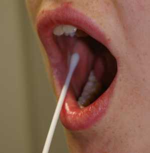
College student Amit Gupta has just under 28 days left to find a suitable bone marrow donor to combat the Leukemia threatening his life by getting people to get a cheek swab to test for compatibility at http://amitguptaneedsyou.com/

cheekswab.jpg

Because South Asians (Indians Pakistanis, Nepali etc.) are so sparsely represented in the donor pool a unique all-out drive has begun to beat the clock and the disease.

“Leukemia is a word no one wants to hear. It is a type of cancer that starts inside your bone marrow. Amit Gupta has it, and his survival is 30% to 35%. A bone marrow match would double his survival. people are severely under reported in the donor pool including other minorities. finding a perfect match are about one in 20,000. Amit has been using social media to get the word out and  reach potential donors. ” Dr.  Sanjay Gupta from CNN, USA.  http://on.cnn.com/sELFzT

The nature of Acute Leukemia is it’s swift and often unheralded, undiagnosed onset. Amit had been feeling worn out and was losing weight, and wasn’t sure why. After an exam and some lab work his doctor called and was brief, “Amit, you’ve got Acute Leukemia. You need to enter treatment right away.” Within hours he was hospitalized and the race had begun.

“I have a couple more months of chemo to go, then the next step is a bone marrow transplant. South Asians are severely under-represented in the bone marrow pool, and I need help,” Amit said.

Unlike blood transfusions, finding a genetic match for bone marrow that his body will accept is no easy task. The national bone marrow registry has 9.5 million records on file, yet the chances of someone from South Asian descent of finding a match are only 1 in 20,000.

Amit started the photo websites Photojojo and Jelly. Both sites have galvanized into action with “Swab for Amit” gatherings, making calls to South Asian clubs and putting out the word all over the Internet.

A few ways to help:

  1. If you’re South Asianget a free test by mail. You rub your cheeks with a cotton swab and mail it back. It’s easy.
  2. Anyone who is South Asian (India, Pakistan, Bangladesh, Nepal, Bhutan, Maldives, or Sri Lanka), please point them to http://amitguptaneedsyou.com/

Students have organized a donor drive by contacting 100kcheeks@gmail.comThey sent kits, flyers, information and made the whole process easy. From NYC to San Francisco from the UK to India strangers are participating to help find a donor and save Amit’s life and eventually help others.

Enhanced by Zemanta

Scientists create jaw bone from stem cells

image

New York: Scientists have created part of the jaw bone for the first time in the lab using human adult stem cells.

The technique, which created the joint, may be useful for other areas of the body.

The Columbia University study appears in Proceedings of the National Academy of Sciences.

The bone which has been created in a lab, by scientists at Columbia University is called the temporomandibular joint (TMJ). This joint which is complex to repair can be damaged by defects at birth, arthritis or injury. Treatment is notoriously difficult as the joint has a complex structure and is difficult to replicate using bone grafts.

Scientists used human stem cells taken from bone marrow which were then planted into a “tissue scaffold” in the shape of a human jaw bone.

The cells were then cultured nutrients found during natural bone development.

Lead researcher Dr Gordana Vunjak-Novakovic said: “The availability of personalised bone grafts engineered from the patient’s own stem cells would revolutionise the way we currently treat these defects.”

She said the new technique could also be applied to other bones in the head and neck, including skull bones and cheek bones, which are similarly difficult to graft.

She stressed that the joint created in the lab was bone only, and did not include other tissue, such as cartilage which the team is also working on.

Obesity speeds up cartilage loss

image

Boston: Obesity, among other factors, is strongly associated with an increased risk of rapid cartilage loss, according to a study published in the August issue of the magazine Radiology.

“We have isolated demographic and MRI-based risk factors for progressive cartilage loss,” said the study’s lead author, Frank W. Roemer, M.D., adjunct associate professor at Boston University and co-director of the Quantitative Imaging Center at the Department of Radiology at Boston University School of Medicine.

“Increased baseline body mass index (BMI) was the only non-MRI-based predictor identified.”

As obesity is one of the few established risk factors for osteoarthritis, it is not surprising that obesity may also precede and predict rapid cartilage loss. Weight loss is probably the most important factor to slow disease progression.
Risk Factors for MRI-detected Rapid Cartilage Loss of the Tibio-femoral Joint over a 30-month Period: the MOST Study.

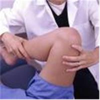
Tibio-femoral cartilage is a flexible connective tissue that covers and protects the bones of the knee. Cartilage damage can occur due to excessive wear and tear, injury, misalignment of the joint or other factors, including osteoarthritis.

Osteoarthritis is the most common form of arthritis, affecting 27 million Americans, according to the National Institute of Arthritis and Musculoskeletal and Skin Diseases. In osteoarthritis, the cartilage breaks down and, in severe cases, can completely wear away, leaving the joint without a cushion. The bones rub together, causing further damage, significant pain and loss of mobility.

The best way to prevent or slow cartilage loss and subsequent disability is to identify risk factors early.

“Osteoarthritis is a slowly progressive disorder, but a minority of patients with hardly any osteoarthritis at first diagnosis exhibit fast disease progression,”

Dr. Roemer said. “So we set out to identify baseline risk factors that might predict rapid cartilage loss in patients with early knee osteoarthritis or at high risk for the disease.”

The researchers recruited patients from the Multicenter Osteoarthritis (MOST) Study, a prospective study of 3,026 people, age 50 – 79, at risk for osteoarthritis or with early x-ray evidence of the disease. The study is funded by the National Institute on Aging.

Dr. Roemer’s study consisted of 347 knees in 336 patients. The patient group was comprised of 65.2 percent women, mean age 61.2, with a mean BMI of 29.5, which is classified as overweight. Recommended BMI typically ranges from 18.5 to 25. Only knees with minimal or no baseline cartilage damage were included. Of 347 knees selected for the study, 20.2 percent exhibited slow cartilage loss over the 30-month follow-up period and 5.8 percent showed rapid cartilage loss. Rapid cartilage loss was defined by a whole organ magnetic imaging score of at least 5, indicating a large full thickness loss of 75 percent in any subregion of the knee during the follow-up period.

The results showed that the top risk factors contributing to rapid cartilage loss were baseline cartilage damage, high BMI, tears or other injury to the meniscus (the cartilage cushion at the knee joint) and severe lesions seen on MRI at the initial exam. Other predictors were synovitis (inflammation of the membrane that lines the joints) and effusion (abnormal build-up of joint fluid).

Excess weight was significantly associated with an increased risk of rapid cartilage loss. For a one-unit increase in BMI, the odds of rapid cartilage loss increased by 11 percent. No other demographic factors–including age, sex and ethnicity–were associated with rapid cartilage loss.

“As obesity is one of the few established risk factors for osteoarthritis, it is not surprising that obesity may also precede and predict rapid cartilage loss,” Dr. Roemer said. “Weight loss is probably the most important factor to slow disease progression.”

AT A GLANCE

* Researchers using MRI have identified risk factors for rapid cartilage loss in the knee.
* People with a high body mass index (BMI) may be at increased risk for rapid cartilage loss and osteoarthritis.
* Osteoarthritis affects 27 million Americans.

“Risk Factors for MRI-detected Rapid Cartilage Loss of the Tibio-femoral Joint over a 30-month Period: the MOST Study.” Collaborating with Dr. Roemer were Yuqing Zhang, D.Sc., Jingbo Niu, M.D., John A. Lynch, Ph.D., Michel D. Crema, M.D., Monica D. Marra, M.D., Michael C. Nevitt, Ph.D., David T. Felson, M.D., M.P.H., Laura Hughes, Georges El-Khoury, M.D., Martin Englund, M.D., Ph.D., and Ali Guermazi, M.D., for MOST study investigators.

Radiology is edited by Herbert Y. Kressel, M.D., Harvard Medical School, Boston, Mass., and owned and published by the Radiological Society of North America, Inc. (http://radiology.rsnajnls.org/)

RSNA is an association of more than 43,000 radiologists, radiation oncologists, medical physicists and related scientists committed to excellence in patient care through education and research. www.RSNA.org

For patient-friendly information on MRI, visit www.RadiologyInfo.org

OSTEOPOROSIS & BONE HEALTH

US company offers storage of stem cells from menstral blood

image

US company Cryo-Cell has launched a bank designed for women who want to store their own stem cells, taken from the menstrual blood, as a future health insurance.

Stem cells can be obtained from numerous sources, including the blood, bone marrow and embryos. And a number of private companies, including Richard Branson’s Virgin Health Bank, already offer umbilical cord blood banking for about £1,500.

Cryo-Cell charges a sum of $499 (£238) for processing and a year’s storage of menstrual stem cells.

The woman is sent a collection kit in the post, comprising a cup, collection tubes and a prepaid return shipment to Cryo-Cell.

Menstrual stem cells – which form in the womb lining whichis then shed during a woman’s period – have the advantage of being easily harvested in a painless, non-invasive manner as compared to some other stem cell sources such as bone marrow.

And like other stem cells, early lab work suggests they too have the potential to turn into many other types of cell, including heart, nerve, bone, cartilage and fat, the company claims.

Spokesman for the company, stem cell expert Dr Stephen Noga, director of the Cellular Therapeutics Program, at Sinai Hospital of Baltimore, said: “Even one menstrual cycle has the potential to produce millions of stem cells.

“Current research is very preliminary, but given their properties, we believe these menstrual stem cells demonstrate compelling promise to transform regenerative medicine in the coming years.”

Cryo-Cell says on its website that “realistically, it may take several years for these menstrual stem cells to be developed into potential widely-available commercial therapies”.

Bones act as an organ – new research reveals

Even though bones seem to be metabolically inactive structures, nothing could be further from the truth. In fact, bones are rebuilt constantly through the action of cells known as osteoblasts while old bone is destroyed by other cells known as osteoclasts. Bones also produce red and white blood cells, help maintain blood pH and store calcium.

However, exciting new research published in this month’s edition of the magazine Cell, has shown that bones also act as an endocrine organ. Not only do bones produce a protein hormone, osteocalcin that regulates bone formation, but this hormone also protects against obesity and glucose intolerance by increasing proliferation of pancreatic beta cells and their subsequent secretion of insulin. Osteocalcin was also found to increase the body’s sensitivity to insulin and as well as reducing its fat stores.

Hormones function as chemical messengers that allow the body to precisely coordinate metabolism, reproduction and other essential biological processes that involve multiple organs.

“The skeleton used to be thought of as just a structural support system. This opens the door to a new way of seeing the bones,” said Dr. Gerard Karsenty, chairman of the department of genetics and development at Columbia University Medical Center in NYC, who headed the team that made the discovery.

Osteocalcin is not new to science: Its existence has been known for 50 years, “but its function was never understood,” observed Karsenty. However, researchers have long known that people with diabetes tend to have low levels of osteocalcin, but until now no one understood the significance.

Based on their knowledge of skeletal biology and endocrinology, the research team hypothesized that there might be a relationship between skeletal biology and endocrine regulation because of the long-known observation that obesity protects against osteoporosis in mammals. Additionally, it was known that people with untreated type 2 diabetes have low osteocalcin levels, which made this hormone an appealing target for their research efforts.

To do this research, the scientists designed an elegant series of experiments using several groups of mice. The first group of experimental mice had their osteoblast gene, called Esp, genetically deactivated, or “knocked out”. Esp encodes a receptor-like protein tyrosine phosphatase called OST-PTP that increases beta-cell proliferation and insulin secretion in the pancreas, which results in hypoglycemia. But these so called “knock-out mice” lacked all functional Esp genes, so their insulin secretion and sensitivity decreased causing them to become obese and then to develop Type 2 diabetes when fed a normal diet. Type 2 diabetes occurs when the body becomes resistant to insulin, the hormone that regulates sugar metabolism.

A second group of experimental mice were genetically engineered to over-produce osteocalcin. These mice showed lower-than-normal blood glucose levels and higher insulin levels than did normal mice that were fed a normal diet. Additionally, these “overproducer mice” also showed increased insulin sensitivity. This is probably the most exciting result because typically, excess blood insulin decreases tissues’ sensitivity to the hormone, which makes insulin treatment difficult for diabetics. Further, the team found that treating the “knock-out mice” with osteocalcin helped regulate their blood sugar and insulin.

Additionally, the investigators reported that mice with one functional copy of Esp showed a significant reversal of their metabolic abnormalities, which provides “genetic evidence that Esp and osteocalcin lie in the same regulatory pathway and that [the] Esp-/- mice metabolic phenotype is caused by a gain-of-activity of this hormone.”

Interestingly, mice that are genetically programmed to overeat and mice that were fed fatty diets were prevented from suffering both obesity and diabetes when given high levels of osteocalcin. Karsenty is now determining whether giving osteocalcin to his diabetic “knock-out mice” will reverse the disease. This research shows promise for treating human diabetics as well.

Finding a substance that increases beta cell proliferation, says Karsenty, “is a holy grail for diabetes research.” Thus, if what’s true for mice also proves true for humans, “then we have inside us a hormone that does precisely this.”

“The findings could have important implications for the treatment of diabetes. Osteocalcin has a triple-punch effect, in that it raises both insulin levels and insulin uptake while keeping fat at bay. That makes it a promising therapy for middle-aged people who want to fight type 2 diabetes,” Karsenty said.

Additionally, this study also reveals that the skeleton is an important part of the endocrine system.

“To our knowledge this study provides the first in vivo evidence that [the] skeleton exerts an endocrine regulation of energy metabolism and thereby may contribute to the onset and severity of metabolic disorders,” the authors wrote in their paper.

Doctors research use of stem cells to grow new bone

York: Doctors in the UK are investigating how bone can be grown from baby stem cells to replace diseased joints.

At present stem cells from the umbilical cords of babies are used to treat leukaemia patients, but researchers at York University hope to find a new use for the two million units of cord blood collected every year in Europe.

Doctors already believe the process is possible but now need clinical proof, which is being funded by the EU to the tune of £1.6 million. Last year doctors in Germany replaced part of the skull of a child using stem cells taken from body fat, which appeared to turn into bone cells.

German doctors grow new jawbone from stemcells

Kiel: A German man was able to chow down on a bratwurst sandwich after surgeons grew a new jaw bone on his back muscle and transplanted the bone to his mouth.

The 56-year-old man had part of his lower jaw removed because of mouth cancer. Until the transplant, his diet was limited to soft food and soup for nine years.

Doctors in Kiel, Germany have rebuilt a man’s face using a new jaw grown on his back.

Dr Patrick Warnke, a reconstructive facial surgeon at the University of Kiel in Germany and his colleagues used CT scans and computer-aided designs to build a virtual jaw replacement.

The researchers used the model to construct a mesh cage, which they filled with bone mineral, protein and stem cells from the patient’s bone marrow. They then transplanted the implant below his right shoulder blade.

Seven weeks later, surgeons transplanted the new bone graft into the mouth gap in the patient’s face.A month after the surgery, he was able to eat his celebratory supper.The experiment is described in the medical journal The Lancet.

“We suggest that our results represent a proof of principle,” Warnke said in a release, noting the patient needs to be followed in the long term before drawing conclusions.

Researchers need to investigate appropriate combinations of biomaterials, growth factors and “blank slate” stem cells needed to build strong, resilient substitutes for bone, said Stan Gronthos, a stem cell expert at the Hanson Institute in Adelaide, Australia.

“A patient who had previously lost his mandible through the result of a destructive tumour can now sit down to chew his first solid meals in nine years … resulting in an improved quality of life for that individual,” Gronthos wrote in a commentary accompanying the study.